1. Khurshid B, Mousa A, Dallas SL, Deering J, Reznikov N, McKee MD.
Trabecular bone formation in vitro by the OmGFP66 osteogenic cell line.
Bone, https://doi.org/10.1016/j.bone.2025.117767.
2. Bromage TG, Denys C, De Jesus CL, Erdjument-Bromage H, Kullmer Schrenk F, McKee MD, Reznikov N, Ittah E, Buss DJ, Ashley GM, Sandrock O, Pudel SB, Yakar S, Hu B, Rabieh S, Neubert TA.
Fossil mammalian bone paleometabolomes yield metabolic and ecologic profiles.
Nature. https://doi.org/10.1038/s41586-025-09843-w.
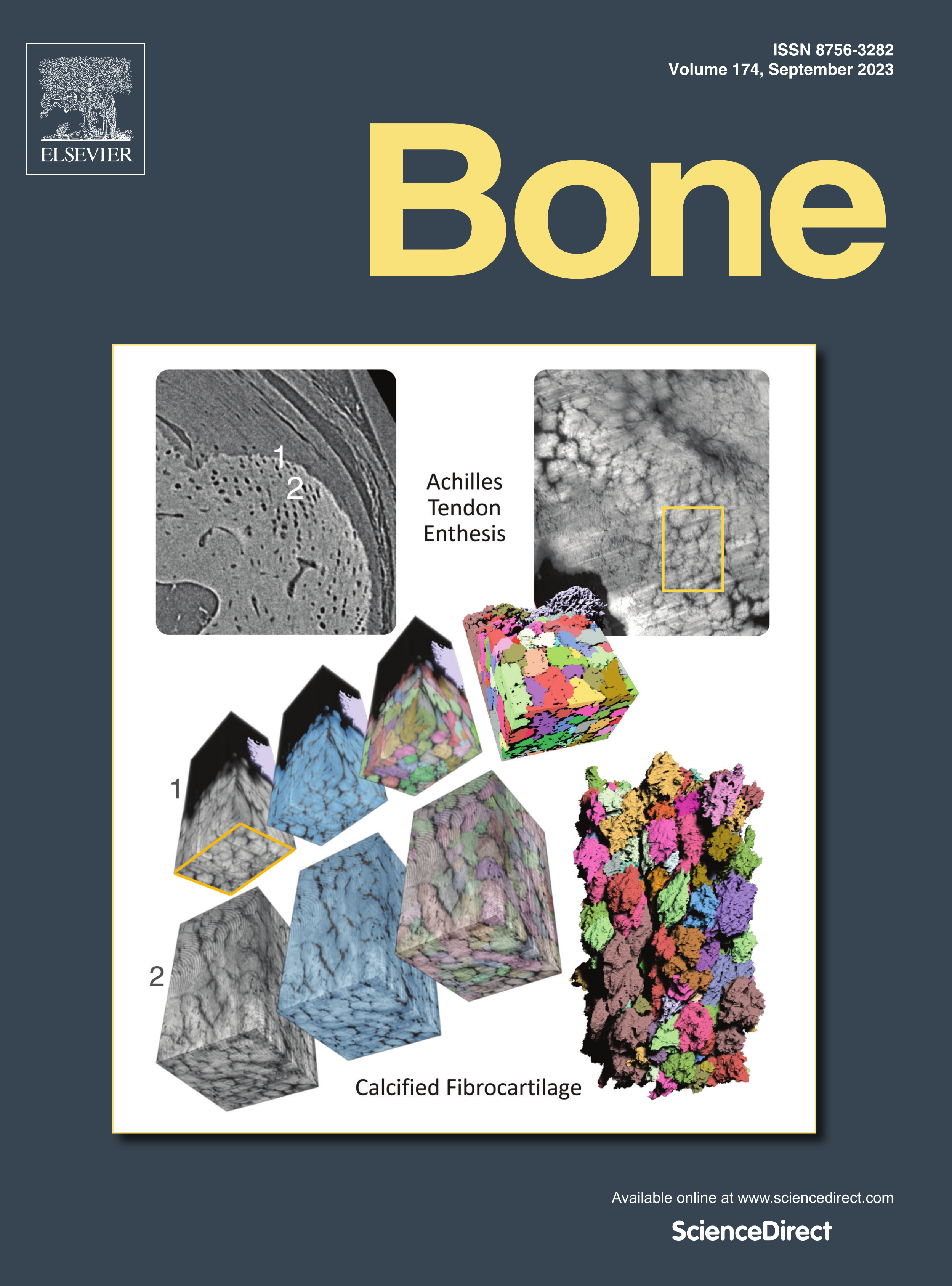
3. Deering J, Buss DJ, Murshed M, Reznikov N, McKee MD.
Local fibril orientation guides mineral growth direction and morphology in collagenous matrices: A study of parallelism in bone, cartilage, enthesis fibrocartilage, calcifying tendon and dentin.
ACS Nano, https://doi.org/10.1021/acsnano.5c11922.
4. Jia S, Piché N, McKee MD, Reznikov N.
Advancing X-ray micro-computed tomography image processing of avian eggshells: An improved registration metric for multiscale 3D images and resolution-enhanced segmentation using edge-attentive neural networks.
Micron. 199:103915. https://doi.org/10.1016/j.micron.2025.103915
5. Rudski BZ, Deering J, Reznikov N.
The Anisotropy Vector Rose: A new package for analysing and visualising 3D non-unit vectors in Python.
J Open Source Software, 2025, 10(111):8369 https://doi.org/10.21105/joss.08369
6. Nelea V, Ittah E, McKee MD, Reznikov N.
Bone mineral tessellation: Visualization by atomic force microscopy of the volume-filling mineralization pattern in hydrated and dehydrated states.
Acta Biomaterialia. 2025 https://doi.org/10.1016/j.actbio.2025.05.016.
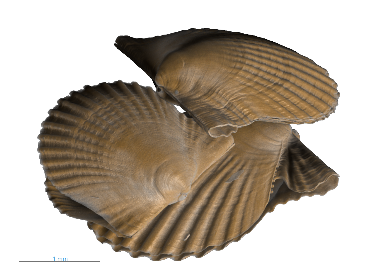
7. Khurshid B, Benchetrite A, Guichaoua L, Brodusch N, Stewart BD, Kröger R, Gauvin R, Mallet M, Tremblay R, Reznikov N
Investigating temperature influences on shell growth and microstructural variations in Bay scallops: Insights from multiscale microscopy
Faraday Discussions 2025 https://doi.org/10.1039/D5FD00023H
8. Guichaoua L, Reznikov N, Stewart BD, Kröger R, Gauvin R
Combined crystallographic study of king scallop (Pecten maximus) shells using SEM, EBSD and Raman spectroscopy
Faraday Discussions 2025 https://doi.org/10.1039/D5FD00029G
9. Deering J, Buss DJ, Kröger R, Vali H, Lagos MJ, Reznikov N, McKee MD
Bone mineralization and the effects of elevated osteopontin: From symmetry-breaking foci to 3D space-filling tessellation
Faraday Discussions 2025 https://doi.org/10.1039/D5FD00013K
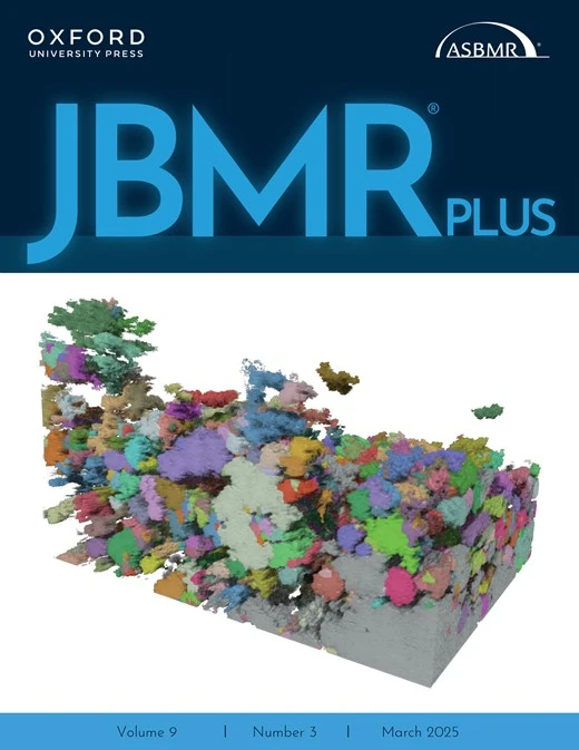
10. Buss DJ, Deering J, Reznikov N, McKee MD
Understanding the structural biology of osteomalacia through multiscale 3D X-ray and electron tomographic imaging: A review of X-linked hypophosphatemia, the Hyp mouse model, and imaging methods.
J Bone Miner Res Plus, 2025 (special issue on rare bone diseases), 9(2): ziae176. https://doi.org/10.1093/jbmrpl/ziae176 (Cover article)
11. Reznikov N
Editorial [for paper of the year 2022] for Buss et al., “Hierarchical organization of bone in three dimensions; a twist of twists”
J Struct Biol: X, 2024(10):100116 http://doi.org/10.1016/j.yjsbx.2024.100116
12. Reznikov N
The Stenciling Principle for extracellular matrix mineralization. Textbook Sub-Chapter in: Ten Cate's Oral Histology: Development, Structure, and Function (10th edition)
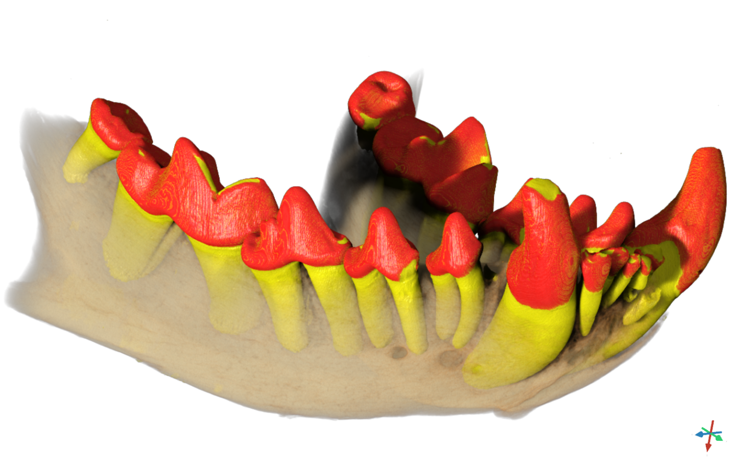
Author/Editor Antonio Nanci. Elsevier. ISBN-13 978-0323798952 https://shop.elsevier.com/books/ten-cates-oral-histology/nanci/978-0-323-79895-2#full-description
13. Halgrain M, Schneider MA, Jia S, Narcy A, Gambier E, Hincke MT, McKee MD, Rehault-Godbert S, Reznikov N
A 3D micro-computed tomography study comparing embryonic skeletal development in layer versus broiler strains of the domestic chicken
Poultry Science, 2024, 103(12):104308 http://doi.org/10.1016/j.psj.2024.104308
14. Rodriguez-Navarro AB, Dominguez-Gasca N, Athanasiadou D, Le Roy N, Reznikov N, Gonzalez-Segura A, Hincke MT, McKee MD, Nys Y, Gautron J
Guinea fowl eggshell structural analysis at different scales reveals how organic matrix induces microstructural shifts that enhance its mechanical properties
Acta Biomater, 2024, 178:244-256 http://doi.org/10.1016/j.actbio.2024.03.001
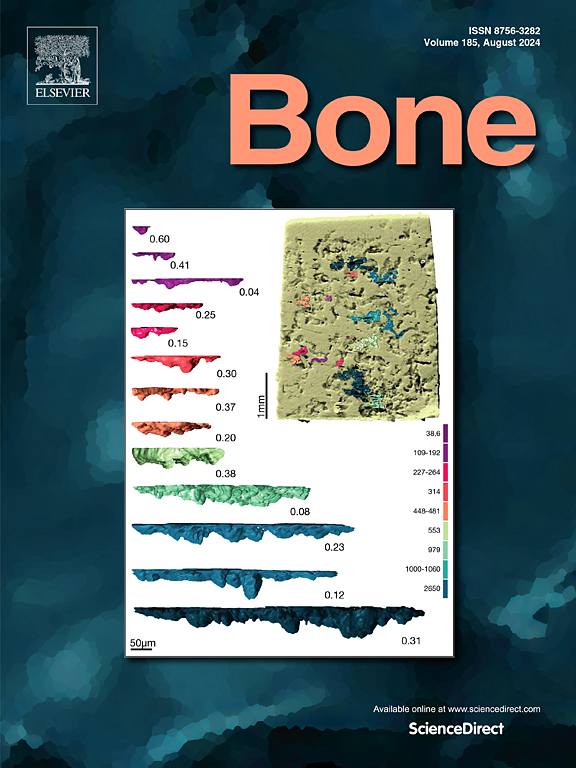
15. Grass D, Malek G, Taieb H, Ittah E, Richard H, Reznikov N, Laverty S
Characterization and quantification of in-vitro equine bone resorption in 3D using microcomputed tomography (µCT) and deep learning-aided feature segmentation
Bone, 2024, 185:117131 https://doi.org/10.1016/j.bone.2024.117131
16. Daniel J Buss, Natalie Reznikov, Marc D McKee
Attaching organic fibers to mineral: The case of the avian eggshell
iScience, 2023 https://doi.org/10.1016%2Fj.isci.2023.108425
17. Daniel J Buss, Katya Rechav, Natalie Reznikov, Marc D McKee
Mineral tessellation in mouse enthesis fibrocartilage, Achilles tendon, and Hyp calcifying enthesopathy: A shared 3D mineralization pattern
Bone, 2023 https://doi.org/10.1016/j.bone.2023.116818
18. McKee MD, Buss DJ, Reznikov N.
Mineral tessellation in bone and the stenciling principle for extracellular matrix mineralization
J Struct Biol, 2022, 214(1):107823 https://doi.org/10.1016/j.jsb.2021.107823
19. Reznikov N, Liang H, McKee MD, Piché N.
Mapping of trabecular bone anisotropy and volume fraction in 3D using µCT images of the human calcaneus
Am J Biol Anthrop, 2022 https://doi.org/10.1002/ajpa.24474
20. Buss DJ, Kroeger R, McKee MD, Reznikov N.
Hierarchical organization of bone in three dimensions: A twist of twists
J Struct Biol: X, 2022(6):100057 https://doi.org/10.1016/j.yjsbx.2021.100057
21. Alsheghri A*, Reznikov N*, Piché N, Gendron M, Tamimi Marino F, Song J.
Optimization of 3D network topology for bioinspired design of stiff and lightweight bone-like structures
Mat Sci Eng: C, 2021, 112010 https://doi.org/10.1016/j.msec.2021.112010 * equally contributing authors.
22. Reznikov N, Buss DJ, Provencher B, McKee MD, Piché N.
Deep learning and 3D imaging in structural biology. Review
J Struct Biol, 2020: 107598 (invited from conference selection, special issue) https://doi.org/10.1016/j.jsb.2020.107598
23. Buss DJ, Reznikov N, McKee MD.
Crossfibrillar mineral tessellation in normal and Hyp mouse bone as revealed by 3D FIB-SEM microscopy
J Struct Biol, 2020: 108603 https://doi.org/10.1016/j.jsb.2020.107603
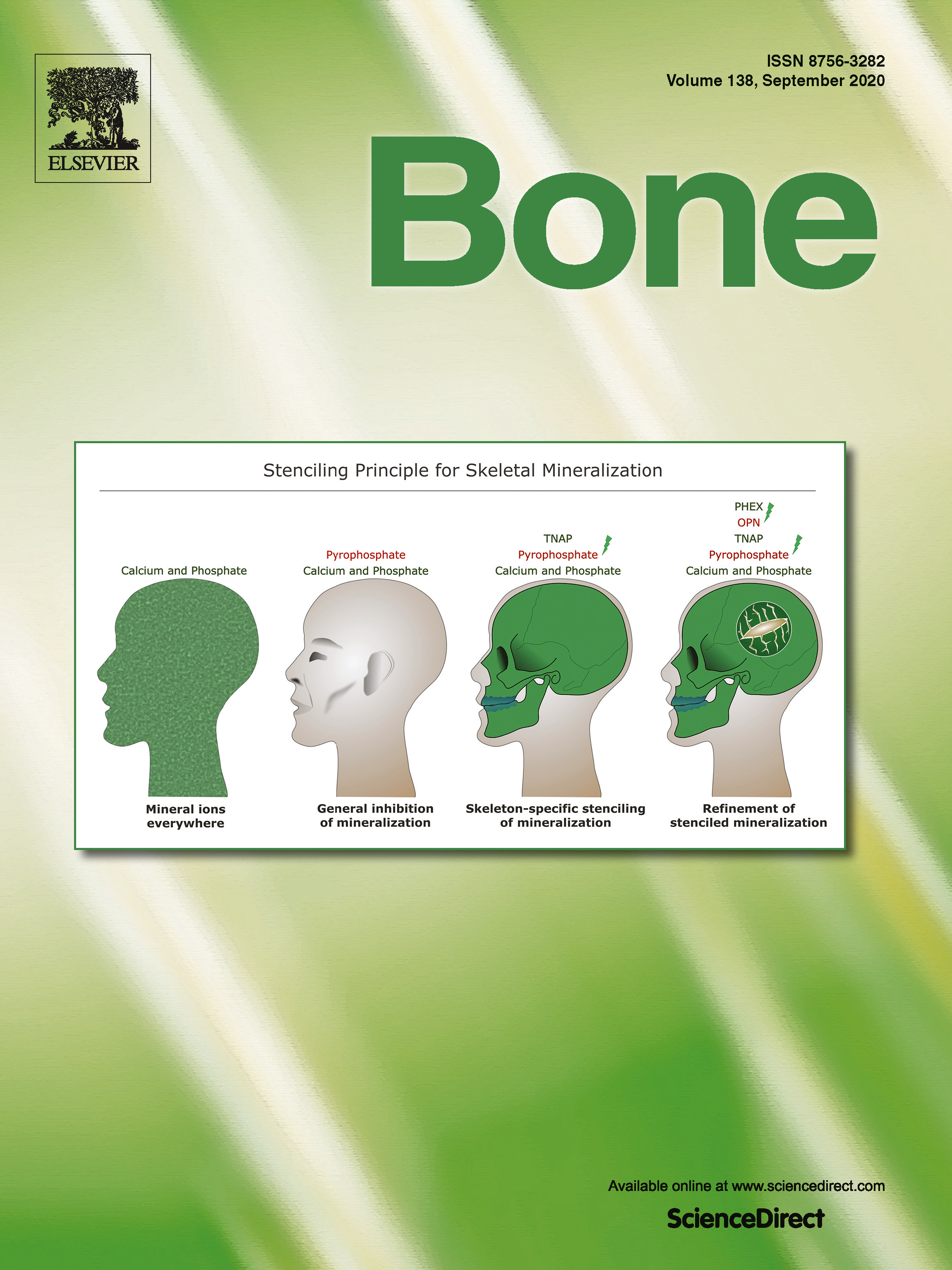
24. Reznikov N, Hoac B, Buss DJ, Addison WN, Barros NMT, McKee MD.
Biological stenciling of mineralization in the skeleton: Local enzymatic removal of inhibitors in the extracellular matrix.
Bone, 2020 https://doi.org/10.1016/j.bone.2020.115447 (cover article)
25. Athanasiadou D, Jiang W, Reznikov N, Rodriguez-Navarro AB, Kroeger R, Bilton M, Nelea V, Hu Y, McKee MD.
Nanostructure of mouse otoconia
J Struct Biol, 2020, 210(2):107489. https://doi.org/10.1016/j.jsb.2020.107489
26. Reznikov N, Rechav K.
FIB-SEM dual-beam microscopy for three-dimensional ultrastructural imaging of skeletal tissues. Methodological insert in Comparative skeletal histology and paleohistology. Editors : A. de Ricqlès, L. Zylberberg, K. Padian and V. de Buffrénil
CRC Press (Francis and Taylor Group) (invited book chapter, in press).
27. Reznikov N, Alsheghri A, Piche N, Gendron M, Morozova I, Sanchez Siles JM, Gonzalez-Quevedo D, Tamimi Marino I, Song J, Tamimi Marino F.
The topological blueprint of trabecular bone associates with skeletal pathology in humans
Bone Reports, 2020 12:100264. https://doi.org/10.1016/j.bonr.2020.100264
28. Reznikov N, Dagdeviren D, Tamimi Marino F, Glorieux F, Rauch F, Retrouvey JM.
Cone-beam computed tomography of Osteogenesis Imperfecta types III and IV: Three-dimensional evaluation of craniofacial features and upper airways
J Bone Miner Res Plus, 2019, 3(6): https://doi.org/10.1002/jbm4.10124
29. Autefage H, Allen F, Tang HM, Kallepitis C, Gentleman E, Reznikov N, Nitiputri K, Nommeots-Nomm A, O’Donnell MD, Lange C, Seidt BM, Kim TB, Lee PD, Pierce BF, Wagermaier W, Fratzl P, Goodship A, Jones JR, Blunn G, Stevens MM.
Multiscale analyses reveal native-like lamellar bone repair and near perfect bone-contact with porous strontium-loaded bioactive glass.
Biomaterials, 2019, 209:152-162. https://doi.org/10.1016/j.biomaterials.2019.03.035
30. Ghouse S, Reznikov N, Boughton O, Babu S, Ng G, Blunn G, Cobb J, Stevens MM, Jeffers J.
The design and in vivo testing of a locally stiffness-matched porous scaffold
Applied Materials Today, 2019, 15:377-388. https://doi.org/10.1016/j.apmt.2019.02.017
31. Reznikov N, Boughton O, Ghouse S, Blunn G, Weston A, Collinson L, Jeffers J, Cobb J, Stevens M.
Individual response variations in scaffold-guided bone regeneration are determined by independent strain- and injury-induced mechanisms
Biomaterials, 2019, 194: 183-194. https://doi.org/10.1016/j.biomaterials.2018.11.026
32. Reznikov N, Bilton M, Lari L, Stevens MM, Kroeger R.
Fractal-like hierarchical organization of bone begins at the atomic level
Science, 2018, 360: eaao2189. https://doi.org/10.1126/science.aao2189
33. Silvent J, Akiva A, Brumfeld V, Reznikov N, Rechav K, Yaniv K, Addadi L, Weiner S.
Embryogenic zebrafish skeleton development: high resolution micro-CT imaging for phenotype identification
PLoS ONE, 2017; 12(12): e0177731 https://doi.org/10.1371/journal.pone.0177731
34. Rowan S, Chang M-L, Reznikov N, Taylor A.
Disassembly of the lens fiber cell nucleus to create a clear lens: The p27 descent
Exper Eye Res, 2017; 156:72-78. https://doi.org/10.1016/j.exer.2016.02.011
35. Ben-Zvi Y*, Reznikov N*, Shahar R, Weiner S.
3D architecture of trabecular bone in the pig mandible and femur: Inter-trabecular angle distributions
Front Mater (Special Issue: Computed Tomography-Based Biomaterials), 2017 https://doi.org/10.3389/fmats.2017.00029
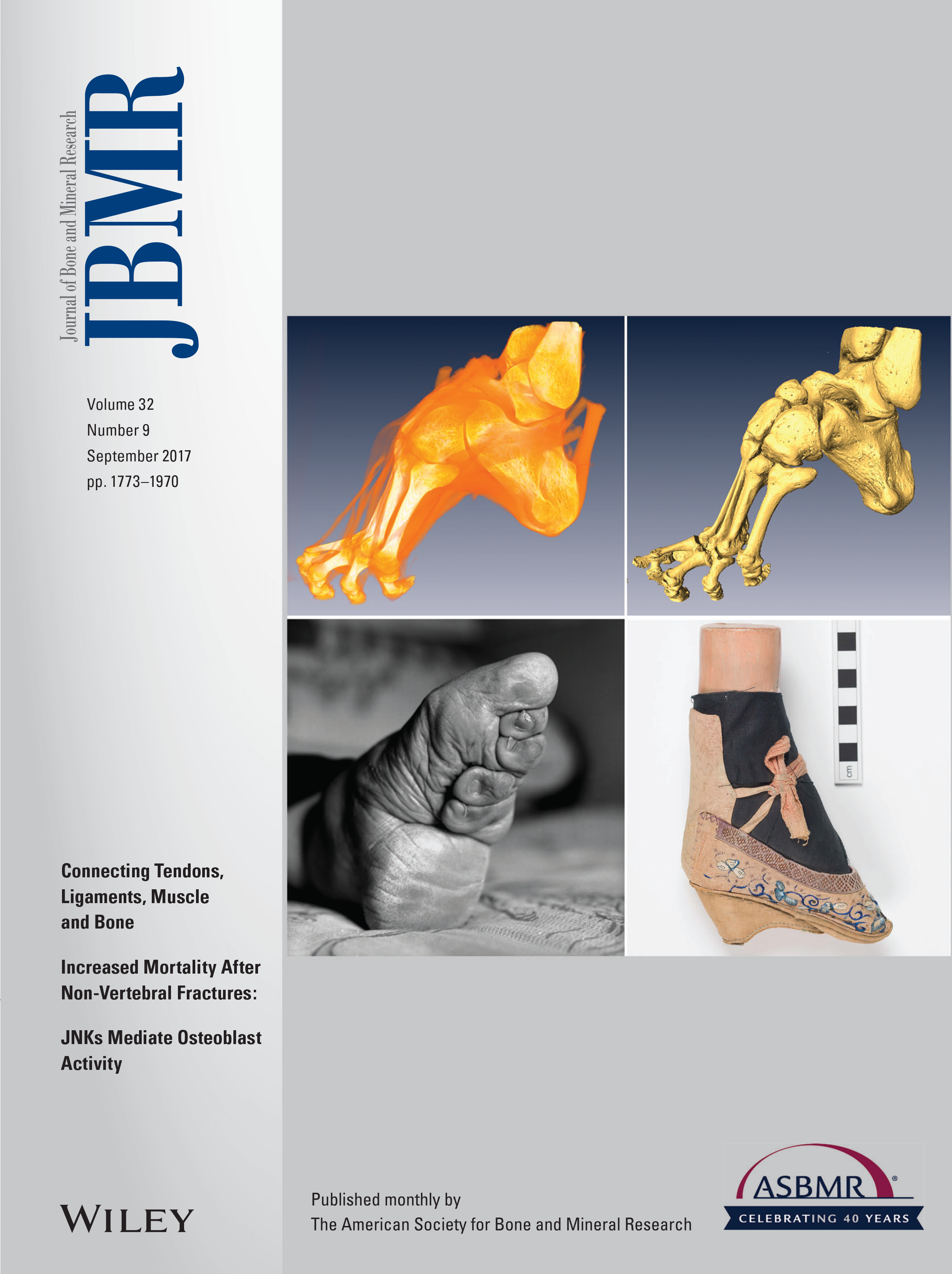
36. Reznikov N, Phillips C, Cooke M, Garbout A, Ahmed F, Stevens MM.
Functional adaptation of the calcaneus in historical foot binding
J Bone Miner Res, 2017, 32(9):1915-1925 https://doi.org/10.1002/jbmr.3185 (cover article, and received American Society for Bone and Mineral Research / Journal of Bone and Mineral Research Raisz-Drezner Award)
37. Kim E, Zwi-Dantsis L, Reznikov N, Hansel CS, Agarwal S, Stevens MM.
One-pot synthesis of multiple protein-encapsulated DNA flowers and their application in intracellular protein delivery
Adv Mater, 2017, 29:1701086 https://doi.org/10.1002/adma.201701086
38. Reznikov N, Steele JAM, Fratzl P, Stevens MM.
A materials science vision of extracellular matrix mineralization
Nature Reviews Mater, 2016:16041. https://doi.org/10.1038/natrevmats.2016.41
39. Reznikov N, Chase H, Ben Zvi Y, Tarle V, Singer M, Brumfeld V, Shahar R, Weiner S.
Intertrabecular angle: a new topological parameter of trabecular bone architecture in the human proximal femur
Acta Biomater, 2016; 44:65-72. https://doi.org/10.1016/j.actbio.2016.08.040
40. Reznikov N, Chase H, Brumfeld V, Shahar R, Weiner S.
The 3D Structure of the collagen fibril network in human trabecular bone: relation to trabecular organization
Bone, 2015; 71:189-195. https://doi.org/10.1016/j.bone.2014.10.017
41. Atkins A, Reznikov N, Ofer L, Masic A, Weiner S, Shahar R.
The three-dimensional structure of anosteocytic lamellated bone of fish
Acta Biomater, 2015; 13:311-323. https://doi.org/10.1016/j.actbio.2014.10.025
42. Reznikov N, Shahar R, Weiner S.
Bone hierarchical structure in three dimensions
Acta Biomater, 2014; 10:3815-3826. https://doi.org/10.1016/j.actbio.2014.05.024
43. Almany-Magal R, Reznikov N, Shahar R, Weiner S.
Three-dimensional structure of minipig fibrolamellar bone: adaptation to axial loading
J Struct Biol, 2014; 186:253-264. https://doi.org/10.1016/j.jsb.2014.03.007
44. Reznikov N, Shahar R, Weiner S.
Three-dimensional structure of human lamellar bone: the presence of two different materials and new insights into the hierarchical organization
Bone, 2014; 59:93-104. https://doi.org/10.1016/j.bone.2013.10.023
45. Reznikov N, Almany-Magal R, Shahar R, Weiner S.
Three-dimensional imaging of collagen fibril organization in rat circumferential lamellar bone using a dual beam electron microscope reveals ordered and disordered sub-lamellar structures
Bone, 2013; 52:676-683. https://doi.org/10.1016/j.bone.2012.10.034
46. Faingold A, Cohen S, Reznikov N, Wagner DH.
Osteonal lamellae elementary units: lamellar microstructure, curvature and mechanical properties
Acta Biomater, 2013; 9:5956-5962. https://doi.org/10.1016/j.actbio.2012.11.032
47. Reznikov N, Barkana I, Abed Y, Har-Zion G, Redlich M.
Measurement of friction forces between stainless steel wires and "reduced-friction" self-ligating brackets
Am J Orth Dent Orthop, 2010; 138:330-338. https://doi.org/10.1016/j.ajodo.2008.07.025
48. Reznikov N, Barkana I, Abed Y, Har-Zion G, Redlich M.
Influence of friction resistance on expression of superelastic properties of initial NiTi wires in "reduced friction" and conventional bracket systems
J Dent Biomech, 2010; 1:613142. http://dx.doi.org/10.4061/2010/613142
Vinay Ashok Kumar
Eran Ittah
Shumeng Jia
Benjamin Rudski
Mahdi Hosseinitabatabaei
Alexandre Demers-Potvin
Alumni
Benazir Khurshid
Lovéni Hanumunthadu
Arnaud Benchetrite
Hubert Taïeb